Welcome to Dolenio doctor sections
To access this section, please complete the details below.
I am a registered UK healthcare professional
Cartilage is 65 to 80 percent water. Three other components make up the rest of cartilage tissue: collagen, proteoglycans, and chondrocytes.
The knees are the body`s primary weight-bearing joints. They are among the joints most commonly affected by osteoarthritis. They may be stiff, swollen, and painful, making it hard to walk, climb, and get in and out of chairs and bathtubs. If not treated, osteoarthritis in the knees can lead to disability. Medications, weight loss, exercise, and walking aids can reduce pain and disability6.
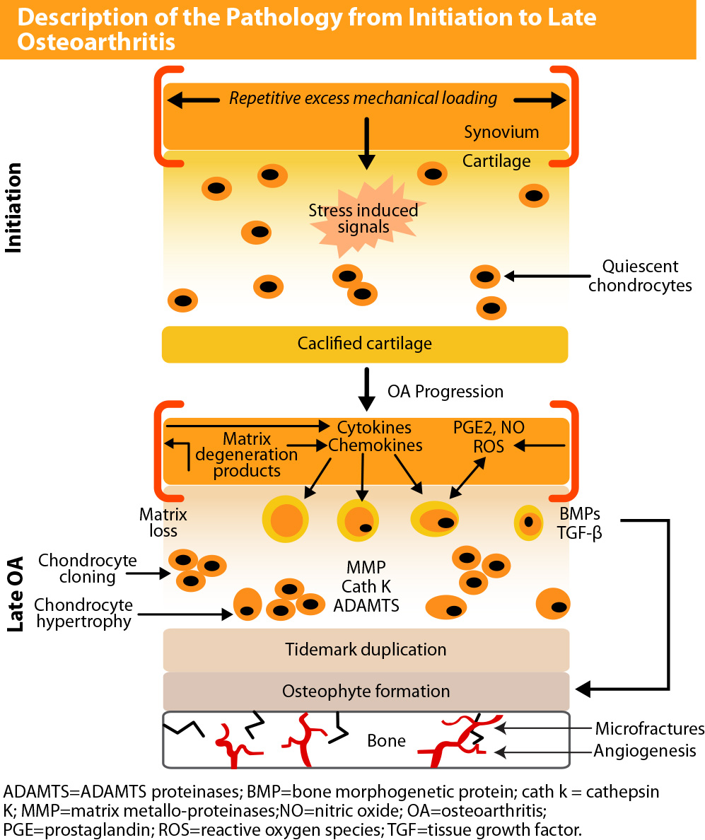
The pathophysiology of OA is complex and can be viewed as the clinical and pathologic outcome of a range of disorders that cause structural and functional failure of the synovial joints with loss and erosion of articular cartilage; subchondral bone alternations;synovial inflammatory responses; and bone and cartilage overgrowth. OA can occur in any of the synovial joints; however, the most common target joint affected is the knee joint.
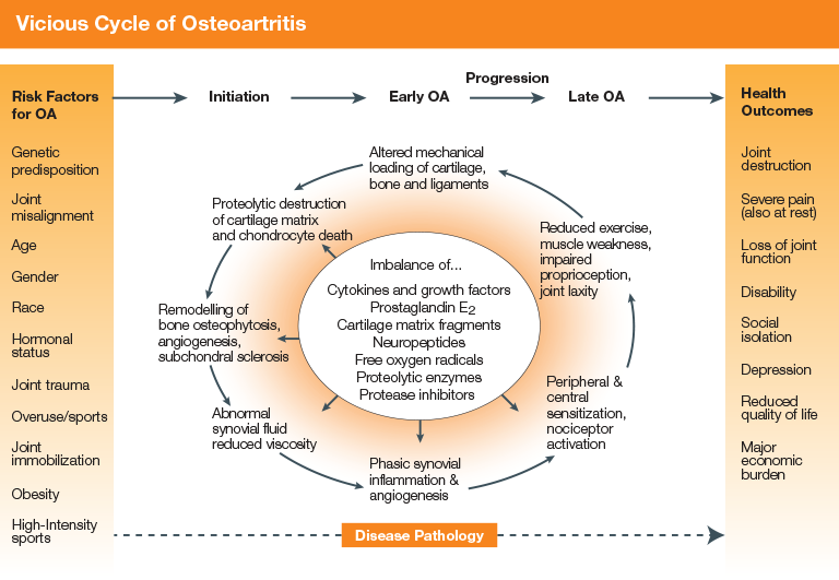
(Figure 1: Source:.
Destruction of cartilage in OA is considered to be a chondrocyte-mediated process. Once initiated, a vicious pathophysiologic process ensues, in which OA may slowly and insidiously progress from early- to late-stage disease, ultimately leading to poor health outcomes7 (figure 1).
In initiation phase, the normally quiescent chondrocytes, as well as the synovial cells, respond to repetitive excess mechanical loading via stress-induced intracellular signals that mediate the production of cytokines, chemokines, other inflammatory mediators (e.g. prostaglandin E2, bradykinin, interleukin [IL], tumor necrosis factora nitric oxide), free oxygen radicals, substance P, and cartilage-degrading proteinases-all of which are extremely toxic and destructive to cartilage
(figure 2).
(Figure 2 : Source:
In an attempt to repair damaged cartilage, chondrocytes undergo proliferation and cloning, which in turn overproduce numerous catabolic and anabolic substances. As cartilage breaks down, it begins to undergo calcification known as tidemark duplication. At the bone–cartilage interface, changes occur in the underlying bone, which thickens with the formation of bony outgrowths (osteophytes) along with the presence of microfactures as a result of invading blood vessels from the subchondral bone. With these blood vessels, sensory nerves also penetrate the area, thus leading to chemical and mechanical stimulation of the nerve endings in these vascular walls. In total, this vicious cycle ends with joint destruction and significant pain burden8.
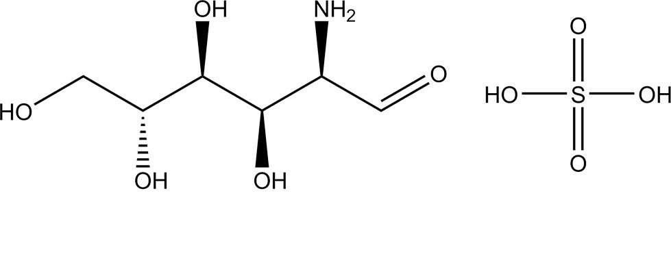
Glucosamine sulphate is a member of glycosaminoglycan family. Glycosaminoglycan is a long chain of amino acids, which is found in high concentration in seashells, from which glucosamine is usually harvested. Glucosamine sulphate is a natural aminosugar comprising glucose, glutamine and sulfur.
Crystalline glucosamine sulfate is a chemically well-characterised pure substance in which glucosamine, sulfate, chloride, and sodium ions are present in stoichiometric ratios of 2:1:2:2, and it is approved as a prescription drug9.